Anterior Cervical Discectomy with Fusion
A Comprehensive Guide by the Brain and Spine Neurosurgical Institute
Introduction to Anterior Cervical Discectomy with Fusion Surgery
If you're grappling with persistent neck and arm discomfort due to nerve root or spinal cord compression, relief may be closer than you think. Our guide provides insights into anterior cervical discectomy with fusion (ACDF), a specialized surgical procedure designed to alleviate symptoms arising from these issues. This comprehensive resource will walk you through the surgery, delve into spine anatomy, outline the procedure's steps, and offer post-operative guidance. Our mission is to empower you with the knowledge needed to make informed decisions about your treatment.
Understanding Spinal Cord Compression and ACDF surgery
The agony of nerve compression causing neck and arm pain is a familiar challenge. ACDF, skillfully performed by our dedicated surgeons, targets the problematic area by removing the problematic disc(s) and part of the bone. The spine is then stabilized with a bone graft and hardware, offering a long-term solution. This procedure is considered when other non-surgical options have been exhausted. Typically, a one-night hospital stay is required.
Anatomy of Spine and Diagnosis
Our visit starts with a thorough examination; patients often present with pain radiating down from their neck out into their arms. This pain is often due to the compression of a nerve. We can identify the source of this pain by looking at an MRI.
Exploring the Vertebra and Nerve Interaction

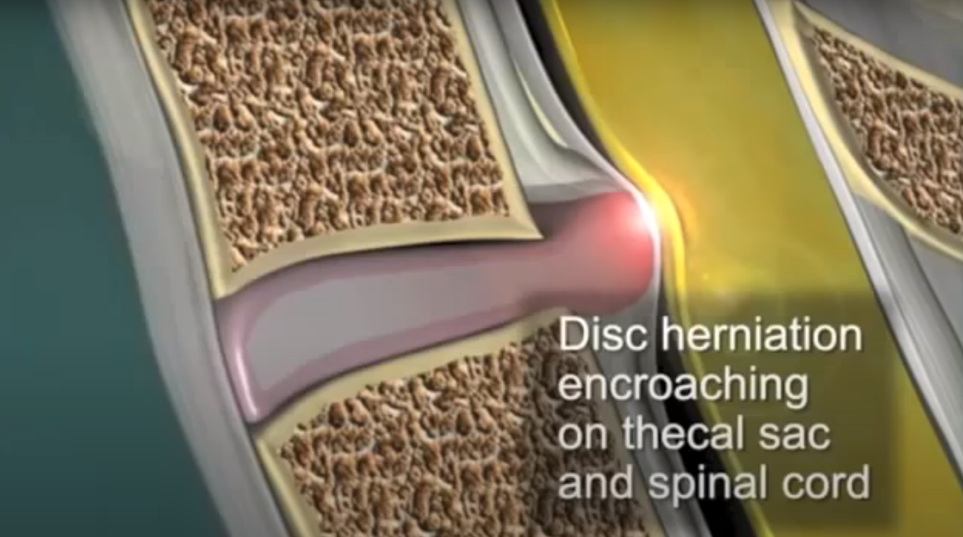
The spine is made up of a group of vertebrae or small bones stacked one on top of the other; these vertebrae are separated by cartilage, a spongy material that provides cushioning between the bones. In the picture above the vertebrae are brown and the cartilage is white. The vertebrae surround and protect the spinal cord which is seen in yellow.

The spinal cord is made up of a collection of nerves and is surrounded by a protective fluid. Above, we see the rapid progression of a herniated disc. The discs of cartilage are constantly supporting our heads and necks. Overtime, this pressure can result in one of these discs being forced out of place and into the spinal canal. As it continues to bulge, this disc may press against a spinal nerve which often results in arm pain.
The ACDF Procedure

In the above MRI, we can see that the patient has a relatively significant disk herniation that is pressing against the spinal column.
The procedure is conducted with the patient lying on his or her back. We can limit the amount of disturbance to the muscles surrounding the spinal cord by operating on the front of the neck. A one to two inch incision is made at the location of the herniated disc. Retractors are then placed to separate the muscles in the neck and gain access to the anterior or frontal portion of the spine. Next, we address the specific location of the disc herniation.
Discectomy and Fusion Process
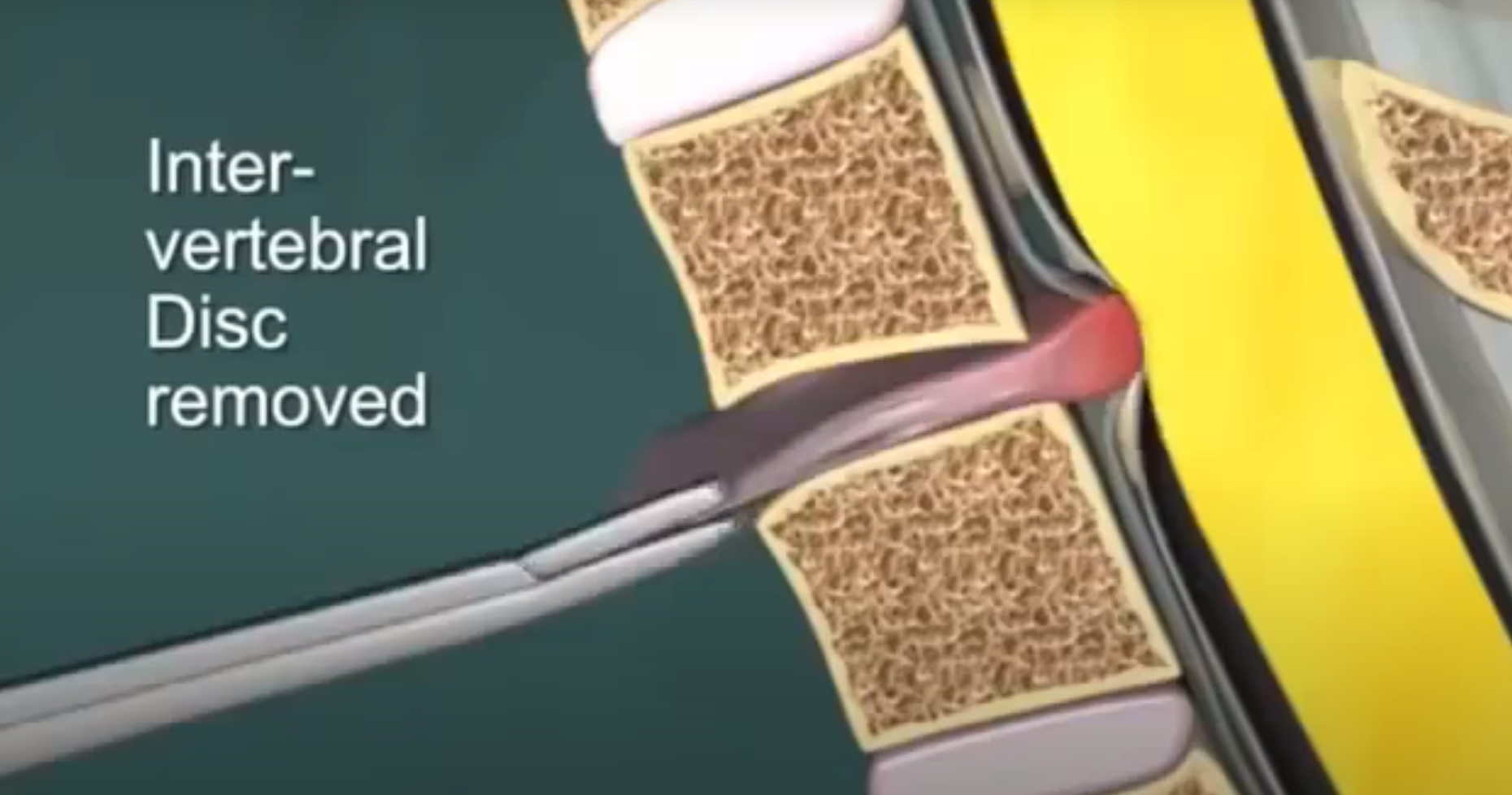
In the photo above, we see a side view of the displaced disc pushing into the spinal canal.
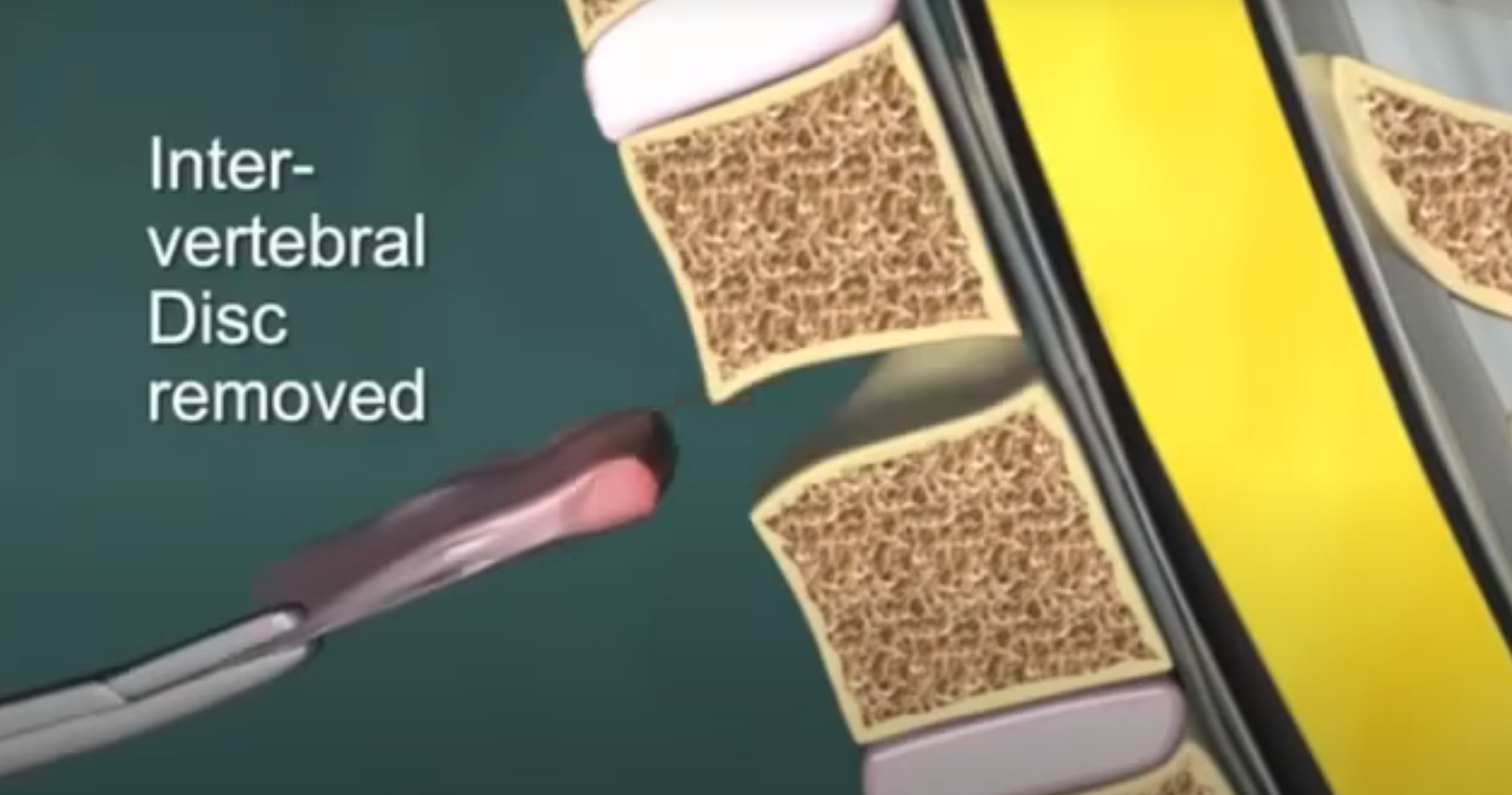
We remove the entirety of the herniated disc; this process is known as a discectomy. With the disk being fully removed, we restructure the spinal canal using a fusion process that promotes the fusion or joining of the vertebra above and below the site of the discectomy.
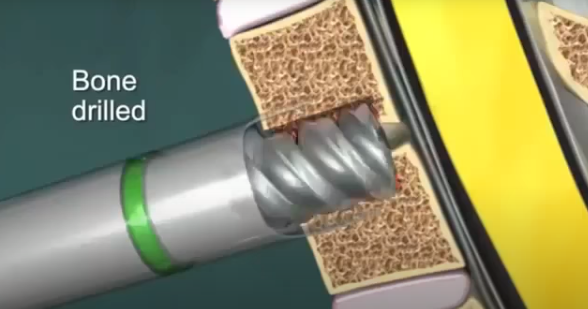
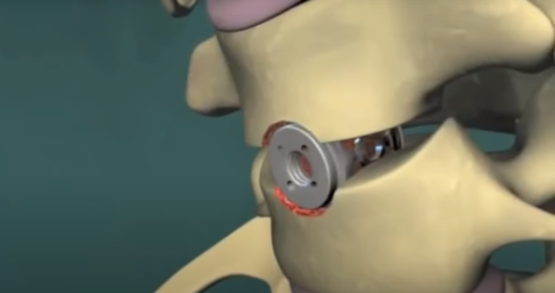
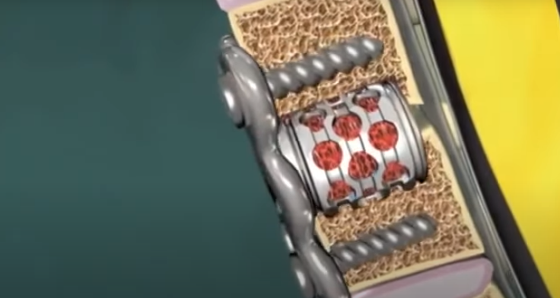
This process begins by drilling into the bone. This is handled very carefully so as not to disturb any surrounding structures. We then place a fusion cage at the site of the discectomy which helps fuse the vertebrae quickly. Small pieces of bone collected during the procedure are then placed around the cage to further aid the fusion. Blades and screws are added to structure the spinal cord during this healing process. Over time the small pieces of bone and the cage will fuse with the vertebra above and below the site of the discectomy. The spinal canal will not be obstructed and any previously experienced pain will be reduced.
Post Surgery
Procedure lasts between one and two hours
Generally completed as an outpatient procedure
Scarring is minimal as the incision is only between one and two inches wide
Patients may experience hoarseness and difficulty swallowing for up to a week after surgery
Arm pain and swelling may present 48 hours after the procedure
Wound Care
Following your surgery, watch our video on correct wound care.
Physical Therapy
After an ACDF operation, we recommend simple exercises. Explore Physical Therapy for Cervical Fusion below.